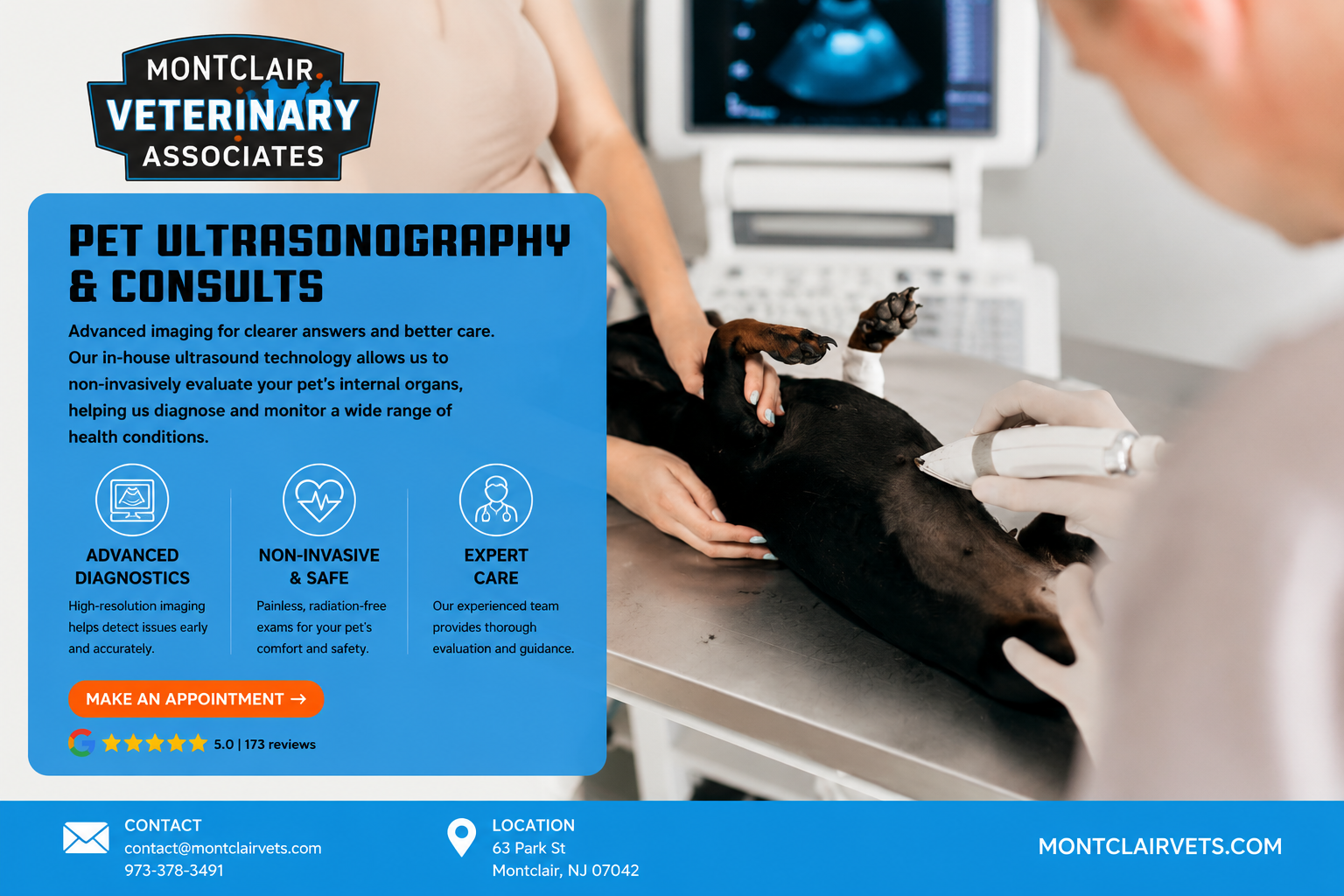
When Is a Pet Ultrasound Recommended?
We may recommend a pet ultrasound when bloodwork reveals abnormal values, when urine test results are inconclusive, or when a mass is felt during a physical exam. Ultrasound allows us to examine internal structures with precision, supporting accurate diagnosis of digestive disorders, urinary issues, endocrine diseases, and more.
In addition to general abdominal imaging, we provide echocardiograms for evaluating heart conditions. Conditions such as heart disease, organ enlargement, and fluid around the pericardial sac can be effectively diagnosed with this approach, often without the need for sedation or general anesthesia.
Advanced Imaging Capabilities and Veterinary Expertise
Our practice is supported by trained ultrasonographers and visiting specialists who bring specialized skill to each exam. We also offer ultrasound-guided fine needle aspiration to sample abnormal tissue, lymph nodes, or internal organs for further testing. These minimally invasive procedures gather diagnostic samples with precision, minimizing discomfort and risk to your pet.
For pets managing chronic illnesses such as kidney disease, liver conditions, or heart disease, repeat ultrasound imaging provides an invaluable window into how a condition is progressing and whether treatment is working as expected.
Safe, Comfortable Procedures Without the Need for Anesthesia
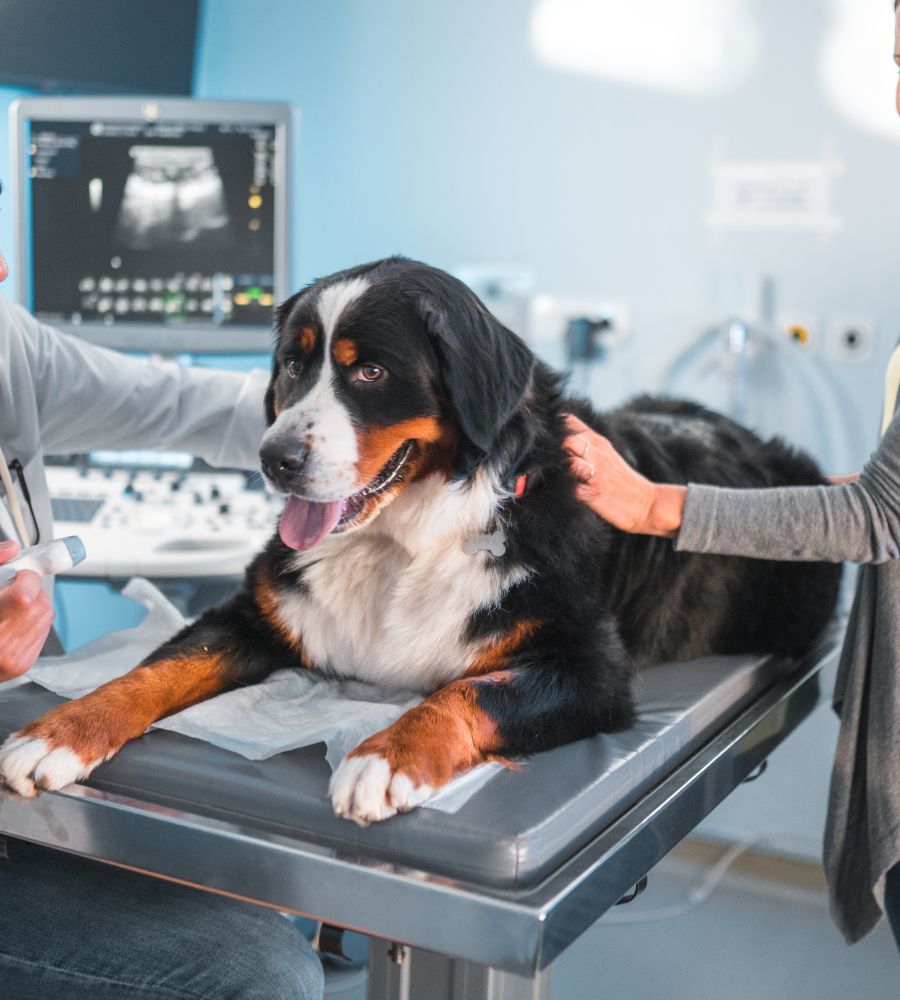
Most ultrasounds can be performed while your pet is awake, resting comfortably on a padded surface. Since it is a non-invasive imaging technique, there is usually no need for general anesthesia unless the patient is particularly anxious or the procedure requires absolute stillness. A small area of fur may be shaved to allow proper contact between the skin and the ultrasound probe.
From start to finish, our veterinary team ensures your pet remains calm and comfortable. Results are typically available the same day, allowing us to make timely decisions about treatment or further testing.
Pregnancy Diagnosis and Reproductive Health
One of the most common uses of ultrasound in veterinary medicine is pregnancy diagnosis. Using real-time imaging, we can monitor fetal development, detect complications, and provide pet owners with peace of mind during a pet’s pregnancy. This is especially helpful in breeding animals or when an accidental pregnancy is suspected.
Ultrasound allows us to visualize individual fetuses, assess their viability, positioning, and growth over time. As a non-invasive and painless option, it is ideal for ongoing monitoring of both the mother and unborn litter without disrupting normal biological processes.
Diagnostic Imaging Services We Provide
At Montclair Veterinary Associates, our full diagnostic imaging suite includes the following services. Each is designed to deliver timely insights with minimal stress to your pet.
- Abdominal ultrasound — including abdominocentesis and cystocentesis procedures
- Thoracic ultrasonography — and thoracocentesis to evaluate the chest cavity
- Doppler ultrasound studies — for assessing blood flow and heart function
- Echocardiogram — for identifying heart disease and structural abnormalities
- Digital radiography (X-ray) — for evaluating bone injuries or lung conditions
Whether we are investigating unexplained symptoms or guiding treatment for a long-term condition, our goal is always to provide an accurate diagnosis and compassionate care.
Pet Ultrasonography Consults at Montclair Veterinary Associates
Dr. Cory Waxman and our team understand that waiting for answers about your pet’s health is stressful. That is why we invest in the equipment and expertise needed to get you those answers quickly. Dr. Waxman earned his degree from the University of Pennsylvania School of Veterinary Medicine and has been treating pets across Northern NJ since 2011, building a reputation for thorough, compassionate diagnostics. To learn more about our team and approach, visit our about page.
If your pet is showing signs of illness, has abnormal lab results, or is due for an imaging evaluation, we are here to help. Contact our office today to schedule a pet ultrasound consult in Montclair, NJ.